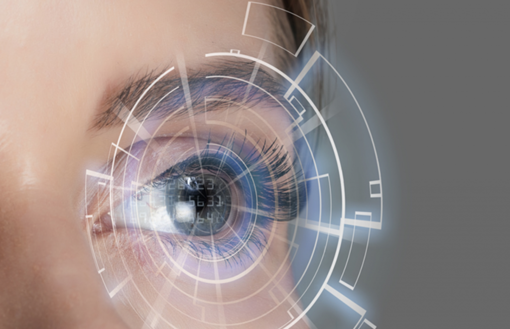
Optometry Technology: Advancing Eye Care
Optometry technology has revolutionized the way we diagnose, treat, and manage eye conditions. From sophisticated imaging techniques to innovative laser surgeries, technology has significantly enhanced the accuracy, efficiency, and comfort […]

Optometry technology has revolutionized the way we diagnose, treat, and manage eye conditions. From sophisticated imaging techniques to innovative laser surgeries, technology has significantly enhanced the accuracy, efficiency, and comfort of eye care. This article explores the fascinating evolution of optometry technology, delving into the advancements that have shaped the field and the exciting possibilities that lie ahead.
The journey of optometry technology began with rudimentary tools and has progressed to encompass a wide range of sophisticated instruments and procedures. These advancements have not only improved patient outcomes but have also opened up new avenues for research and development. From the early days of manual refraction to the advent of automated autorefractors, technology has consistently pushed the boundaries of what is possible in eye care.
Evolution of Optometry Technology

The field of optometry has witnessed a remarkable evolution, driven by technological advancements that have revolutionized the way eye care is delivered. From the rudimentary tools of the past to the sophisticated instruments of today, technological innovation has played a pivotal role in enhancing the accuracy, efficiency, and patient comfort of eye examinations and treatments.
Traditional Methods and Modern Advancements in Eye Examinations
Traditional methods of eye examinations relied heavily on subjective assessments and manual instruments. The Snellen eye chart, for example, remains a fundamental tool for assessing visual acuity, but its limitations in providing comprehensive information about the eye’s health have led to the development of more sophisticated technologies.
- Auto-refractors: These automated devices objectively measure the refractive error of the eye, providing a more accurate and efficient assessment than traditional methods. Auto-refractors use infrared light to determine the shape of the cornea and lens, allowing for precise measurement of refractive error.
- Optical Coherence Tomography (OCT): OCT is a non-invasive imaging technique that provides detailed, cross-sectional images of the retina, optic nerve, and other ocular structures. This technology allows for early detection and diagnosis of various eye conditions, such as glaucoma, macular degeneration, and diabetic retinopathy.
- Fundus Cameras: Fundus cameras capture high-resolution images of the back of the eye, including the retina and optic nerve. These images help optometrists to diagnose and monitor eye conditions, such as diabetic retinopathy and glaucoma.
Technological Innovations in Improving Accuracy, Efficiency, and Patient Comfort
Technological innovations have not only enhanced the accuracy and efficiency of eye examinations but also significantly improved patient comfort.
- Digital Retinoscopy: Digital retinoscopy is a computerized method of measuring refractive error that eliminates the need for manual adjustments and provides a more accurate assessment. This technology is particularly beneficial for children and patients with challenging refractive errors.
- Automated Perimetry: Automated perimetry is a computer-based test that measures peripheral vision, helping to diagnose and monitor conditions such as glaucoma. This technology provides a more accurate and consistent assessment than traditional manual perimetry.
- Contact Lens Fitting Software: Contact lens fitting software uses advanced algorithms to calculate the optimal contact lens parameters for each patient, minimizing trial and error and improving patient comfort. This software also allows for personalized lens designs based on individual eye shapes and refractive errors.
Diagnostic and Imaging Technologies
Optometry relies on a range of diagnostic and imaging technologies to assess eye health, diagnose conditions, and monitor treatment progress. These technologies provide valuable insights into the structure and function of the eye, allowing optometrists to make informed decisions about patient care.
Autorefractors
Autorefractors are automated devices that measure the refractive error of the eye, which is the ability of the eye to focus light onto the retina. This technology utilizes infrared light to measure the eye’s refractive power and determine the appropriate lens prescription.
The principle behind autorefractors is based on the concept of retinoscopy. Retinoscopy involves shining a beam of light into the eye and observing the reflection of the light on the retina. The direction and movement of the reflected light indicate the refractive error of the eye. Autorefractors automate this process, providing a quick and objective assessment of refractive error.
Autorefractors are widely used in optometry for initial eye exams, especially for children and patients who may have difficulty cooperating with traditional subjective refraction. They are also helpful in detecting refractive changes over time, which can indicate the need for updated prescriptions.
Optical Coherence Tomography (OCT), Optometry technology
Optical coherence tomography (OCT) is a non-invasive imaging technique that produces high-resolution, cross-sectional images of the retina and optic nerve. It uses light waves to create detailed images of the different layers of the eye, providing valuable information for diagnosing and managing a wide range of eye conditions.
The principle behind OCT is based on the interference of light waves. A beam of light is directed into the eye, and the reflected light waves are analyzed to create an image. The different layers of the eye reflect light at different depths, allowing OCT to create a detailed cross-sectional view.
OCT has revolutionized the diagnosis and management of various eye conditions, including:
- Macular degeneration: OCT can detect early signs of macular degeneration, allowing for timely intervention and potentially slowing the progression of the disease.
- Diabetic retinopathy: OCT helps monitor the development and progression of diabetic retinopathy, enabling early treatment and preventing vision loss.
- Glaucoma: OCT can assess the thickness of the retinal nerve fiber layer (RNFL), a key indicator of glaucoma damage, aiding in early detection and monitoring of the disease.
- Retinal detachments: OCT can visualize retinal detachments and other retinal tears, facilitating prompt treatment and reducing the risk of vision loss.
Fundus Cameras
Fundus cameras are specialized cameras used to capture high-resolution images of the back of the eye, known as the fundus. These images provide valuable information about the health of the retina, optic nerve, and blood vessels, aiding in the diagnosis and management of various eye conditions.
Fundus cameras use flash photography to illuminate the fundus and capture images. The camera typically uses a red-free filter to enhance the visibility of blood vessels and other retinal structures.
Fundus cameras are essential tools in optometry for:
- Screening for eye diseases: Fundus photography can detect early signs of eye diseases, such as diabetic retinopathy, glaucoma, and macular degeneration.
- Monitoring treatment progress: Fundus images can track changes in the retina over time, providing valuable information about the effectiveness of treatment for various eye conditions.
- Documenting eye conditions: Fundus photographs provide a permanent record of the patient’s eye health, which can be used for future reference and comparison.
Comparison of Imaging Modalities
| Imaging Modality | Advantages | Limitations |
|---|---|---|
| Autorefractors |
|
|
| OCT |
|
|
| Fundus Cameras |
|
|
Refractive Surgery and Laser Technology
Refractive surgery has revolutionized vision correction, offering a permanent alternative to eyeglasses and contact lenses. It involves reshaping the cornea, the clear front part of the eye, to improve its ability to focus light onto the retina. Laser technology plays a crucial role in refractive surgery, enabling precise and controlled corneal reshaping. This section explores the most common refractive surgery techniques, the advancements in laser technology, and the impact on patient outcomes.
Types of Refractive Surgery
Refractive surgery encompasses various techniques, each tailored to specific vision problems. The most common procedures include:
- LASIK (Laser-Assisted In Situ Keratomileusis): LASIK is a widely performed procedure that involves creating a thin flap in the cornea, lifting it, and then using an excimer laser to reshape the underlying corneal tissue. The flap is then repositioned, and it heals naturally. LASIK is effective for correcting myopia (nearsightedness), hyperopia (farsightedness), and astigmatism (irregular curvature of the cornea).
- PRK (Photorefractive Keratectomy): PRK is another laser-based procedure that involves removing the outer layer of the cornea (epithelium) before using an excimer laser to reshape the underlying corneal tissue. The epithelium regenerates over time, typically within a few days. PRK is suitable for individuals with thinner corneas or those who are not candidates for LASIK.
- ICL (Implantable Collamer Lens): ICL is a minimally invasive procedure that involves implanting a small, flexible lens called a phakic IOL (intraocular lens) inside the eye, in front of the natural lens. This lens adds focusing power to the eye, correcting myopia and astigmatism. ICL is an option for individuals with high myopia or those who are not suitable for LASIK or PRK.
Advancements in Laser Technology
Laser technology has significantly evolved since the introduction of refractive surgery, leading to enhanced precision, faster recovery times, and improved patient outcomes.
- Femtosecond Lasers: Femtosecond lasers have replaced mechanical microkeratomes for flap creation in LASIK. These lasers deliver ultra-short pulses of laser energy, allowing for precise and controlled incisions with minimal heat generation. This results in smoother, more predictable flaps and reduced risk of complications.
- Wavefront-Guided LASIK: Wavefront technology analyzes the unique imperfections in a patient’s vision and guides the laser to correct them with greater precision. This personalized approach leads to improved visual acuity and reduced risk of visual aberrations.
- Customizable Ablation Profiles: Modern laser systems allow for customized ablation profiles, tailoring the treatment to each patient’s specific refractive error and corneal anatomy. This personalized approach optimizes visual outcomes and minimizes the risk of under- or over-correction.
Successful Refractive Surgery Cases
Numerous studies and real-world cases have demonstrated the effectiveness and safety of refractive surgery. For example, a study published in the Journal of Refractive Surgery found that 95% of LASIK patients achieved 20/20 or better vision.
“Refractive surgery has been a game-changer for millions of people around the world, offering them freedom from glasses and contact lenses. The advancements in laser technology have made the procedure safer, more effective, and more accessible.”
While refractive surgery is generally safe and effective, potential risks and complications are possible. These can include:
- Under- or Over-correction: This can result in blurred vision that may require additional procedures.
- Dry Eye: Refractive surgery can sometimes affect the tear film, leading to dry eye symptoms.
- Infection: While rare, infection can occur after surgery, requiring treatment with antibiotics.
- Flap Complications: In LASIK, complications related to the flap, such as a flap dislocation or a flap that does not heal properly, can occur.
It’s crucial to consult with a qualified ophthalmologist to determine if refractive surgery is right for you and to understand the potential benefits and risks.
Telemedicine and Remote Eye Care
Telemedicine, the delivery of healthcare services remotely using technology, has revolutionized the field of optometry, offering innovative ways to expand access to eye care and improve patient outcomes. This section delves into the role of telemedicine in optometry, exploring its applications, benefits, and challenges.
Expanding Access to Optometric Services
Telemedicine has the potential to bridge the gap in access to quality eye care, particularly in underserved areas where ophthalmologists and optometrists are scarce. By leveraging technology, telemedicine enables patients in remote locations to connect with qualified eye care professionals for consultations, diagnosis, and monitoring.
- Increased Reach: Telemedicine allows optometrists to reach patients in rural and underserved areas, where traditional healthcare infrastructure may be limited. This is particularly beneficial for individuals who may face barriers to accessing in-person care, such as transportation challenges, financial constraints, or limited availability of specialists.
- Enhanced Accessibility: Telemedicine provides flexibility and convenience for patients, allowing them to consult with eye care professionals from the comfort of their homes. This is especially helpful for individuals with mobility issues, busy schedules, or those who live far from specialized eye care centers.
- Reduced Travel Costs: Telemedicine eliminates the need for patients to travel long distances for appointments, saving them time, money, and effort. This is particularly relevant for individuals who may have to travel extensively for specialized eye care.
Technology Facilitating Remote Eye Examinations
Telemedicine utilizes a range of technologies to facilitate remote eye examinations, consultations, and monitoring. These technologies enable eye care professionals to gather essential information and assess patients’ visual health remotely.
- Teleophthalmoscopy: This technology uses a specialized device to capture images of the retina, allowing optometrists to assess the health of the back of the eye remotely. The images are transmitted to the optometrist’s computer, enabling them to diagnose and monitor eye conditions.
- Remote Visual Acuity Testing: Telemedicine platforms can incorporate visual acuity tests that patients can perform themselves at home. These tests measure the patient’s ability to see clearly at different distances, providing valuable information about their visual acuity.
- Remote Refraction: Telemedicine allows for remote refraction, the process of determining the appropriate prescription for eyeglasses or contact lenses. This involves using specialized equipment and software to measure the patient’s eye’s refractive error remotely.
- Patient Monitoring Devices: Wearable devices and smartphone apps can be used to monitor patients’ eye health remotely. These devices collect data on factors such as eye pressure, visual field, and pupil size, providing valuable insights into potential eye conditions.
The Future of Optometry Technology
The field of optometry is constantly evolving, driven by technological advancements that are revolutionizing eye care. From artificial intelligence to nanotechnology, the future holds exciting possibilities for improving patient outcomes and enhancing the overall experience of vision care.
Emerging Trends and Technological Advancements
The future of optometry is likely to be shaped by several emerging trends and technological advancements.
- Artificial Intelligence (AI) and Machine Learning: AI algorithms can analyze vast amounts of patient data, including medical history, imaging scans, and genetic information, to predict eye disease risk, personalize treatment plans, and improve diagnostic accuracy. For instance, AI-powered systems are being developed to detect early signs of diabetic retinopathy and glaucoma, enabling timely intervention and potentially preventing vision loss.
- Nanotechnology: Nanotechnology is paving the way for innovative ophthalmic devices and treatments. Nanomaterials are being used to create advanced contact lenses with enhanced oxygen permeability, UV protection, and even drug delivery capabilities. Nanoparticles are also being investigated for targeted drug delivery in treating eye diseases like macular degeneration and retinal detachment.
- Bio-inspired Materials: Researchers are exploring bio-inspired materials, such as those found in the human eye, to develop new contact lenses, intraocular lenses, and other ophthalmic devices. For example, researchers are studying the structure of the cornea to develop biocompatible materials for corneal implants, potentially restoring vision in patients with corneal damage.
- Virtual Reality (VR) and Augmented Reality (AR): VR and AR technologies are finding applications in optometry for patient education, visual rehabilitation, and even surgical training. VR simulations can help patients understand their eye conditions and treatment options, while AR can assist surgeons in performing complex procedures with greater precision.
- 3D Printing: 3D printing is revolutionizing the production of customized ophthalmic devices, such as contact lenses, intraocular lenses, and even corneal implants. 3D printed devices can be tailored to the unique anatomy of each patient, improving fit, comfort, and effectiveness.
Impact of Nanotechnology, Gene Therapy, and Bio-inspired Materials on Eye Care
The potential impact of these emerging technologies on eye care is significant, offering hope for treating currently incurable diseases and improving the quality of life for millions of people.
- Nanotechnology: Nanotechnology holds immense potential for delivering drugs directly to the eye, overcoming the limitations of traditional methods. For example, nanoparticles can be designed to target specific cells or tissues in the eye, reducing side effects and improving treatment efficacy. This could revolutionize the treatment of macular degeneration, diabetic retinopathy, and other eye diseases.
- Gene Therapy: Gene therapy has the potential to cure genetic eye diseases by replacing faulty genes with healthy ones. This technology has shown promising results in treating inherited retinal diseases, such as Leber’s congenital amaurosis and retinitis pigmentosa, offering hope for restoring vision in patients who were previously destined to blindness.
- Bio-inspired Materials: Bio-inspired materials are being used to develop artificial corneas, retinal implants, and other biocompatible devices that can restore vision. These materials mimic the natural structure and function of the eye, minimizing rejection by the body and improving long-term success rates. For example, researchers are developing biocompatible materials that can be used to create artificial corneas, potentially restoring vision in patients with corneal damage.
Potential Benefits and Challenges of Future Technologies in Optometry
The adoption of these future technologies in optometry presents both benefits and challenges.
| Benefits | Challenges |
|---|---|
| Improved diagnostic accuracy and early detection of eye diseases | High cost of development and implementation |
| Personalized treatment plans tailored to individual patient needs | Ethical concerns related to data privacy and security |
| Non-invasive and minimally invasive treatment options | Potential for technological failures and complications |
| Enhanced patient education and engagement | Need for ongoing training and education for optometrists |
| Increased accessibility and affordability of eye care | Potential for widening the gap between those who have access to these technologies and those who do not |
Final Thoughts: Optometry Technology
The future of optometry technology holds immense promise, with advancements in artificial intelligence, nanotechnology, and gene therapy poised to further revolutionize the field. These innovations have the potential to personalize treatment plans, enhance diagnostic accuracy, and even prevent or cure eye diseases. As technology continues to evolve, optometrists will be at the forefront of delivering cutting-edge eye care, ensuring that patients receive the best possible treatment and vision outcomes.
Optometry technology has advanced significantly, with digital retinal imaging and automated refraction becoming commonplace. However, as with any technology, there’s a risk of errors and omissions, which can lead to misdiagnosis or treatment complications. Understanding the differences between technology errors and omissions and cyber risks, as outlined in this article technology errors and omissions vs cyber , is crucial for optometrists to mitigate potential risks and ensure patient safety.


