Ophthalmology Technology: Advancing Eye Care
Ophthalmology technology is revolutionizing the way we diagnose, treat, and prevent eye diseases. From sophisticated imaging devices to minimally invasive surgical techniques, these advancements are improving patient outcomes and expanding […]

Ophthalmology technology is revolutionizing the way we diagnose, treat, and prevent eye diseases. From sophisticated imaging devices to minimally invasive surgical techniques, these advancements are improving patient outcomes and expanding access to eye care services.
The field has witnessed a remarkable evolution, transitioning from traditional methods to modern, technologically advanced approaches. This journey has been marked by key breakthroughs that have significantly impacted patient care.
Evolution of Ophthalmology Technology
The field of ophthalmology has witnessed a remarkable transformation, driven by technological advancements that have revolutionized the diagnosis, treatment, and management of eye conditions. From rudimentary tools to sophisticated instruments, ophthalmology technology has played a pivotal role in improving patient care and enhancing visual outcomes.
Early Innovations and Their Impact
The evolution of ophthalmology technology began with the invention of the eye chart in the 18th century, which provided a standardized method for assessing visual acuity. The development of the ophthalmoscope in the 19th century allowed doctors to visualize the interior of the eye, leading to significant advancements in the diagnosis and treatment of eye diseases. Early microscopes and magnifying glasses enabled ophthalmologists to examine the eye in greater detail. These early innovations laid the foundation for the development of more sophisticated technologies in the years to come.
Transition to Modern Technology
The 20th century marked a significant shift in ophthalmology technology, with the introduction of lasers, ultrasound imaging, and other advanced techniques. Lasers revolutionized the treatment of various eye conditions, including glaucoma, diabetic retinopathy, and cataracts. Ultrasound imaging provided a non-invasive way to visualize the eye’s internal structures, while fluorescein angiography allowed doctors to examine the blood vessels in the retina. The development of intraocular lenses (IOLs) for cataract surgery significantly improved visual outcomes for patients.
Comparison of Older and Current Technologies
The capabilities of older technologies have been surpassed by modern advancements in ophthalmology. For example, traditional methods for treating glaucoma, such as medications and surgery, have been complemented by laser procedures and minimally invasive techniques that offer greater precision and efficacy. Ultrasound imaging has evolved into advanced modalities like optical coherence tomography (OCT), which provides detailed, high-resolution images of the retina and other eye structures. Similarly, the use of fluorescein angiography has been largely replaced by OCT angiography, which offers a more accurate and non-invasive way to visualize blood flow in the eye.
Key Breakthroughs and Their Impact
- Laser Technology: The development of lasers in the 1960s revolutionized ophthalmology. Lasers enabled precise and minimally invasive procedures for various eye conditions, such as glaucoma, diabetic retinopathy, and cataracts. The use of lasers for refractive surgery, such as LASIK, has significantly improved vision for millions of people worldwide.
- Optical Coherence Tomography (OCT): Introduced in the 1990s, OCT has become a mainstay in ophthalmology, providing detailed cross-sectional images of the retina and other eye structures. This technology has revolutionized the diagnosis and monitoring of various eye diseases, including glaucoma, macular degeneration, and diabetic retinopathy.
- Intraocular Lenses (IOLs): The development of IOLs in the 1960s revolutionized cataract surgery. IOLs are artificial lenses implanted in the eye to replace the natural lens that has become cloudy due to cataracts. Advancements in IOL technology have resulted in lenses with improved clarity, durability, and multifocality, providing patients with excellent visual outcomes.
Diagnostic Tools and Techniques: Ophthalmology Technology

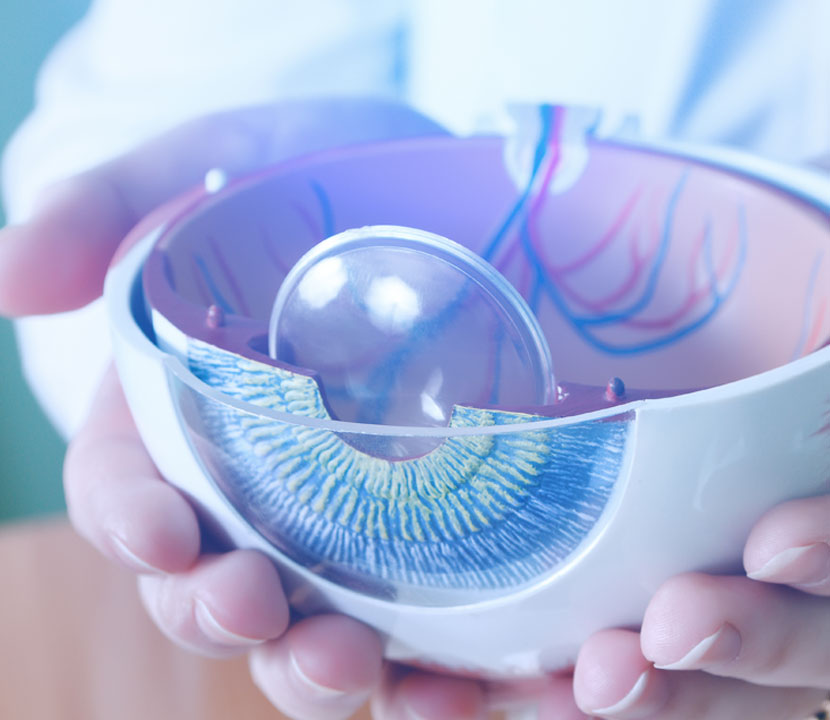
Ophthalmology, the branch of medicine focusing on the eye, has witnessed significant advancements in diagnostic tools and techniques. These advancements have significantly improved the accuracy and efficiency of eye disease diagnosis, leading to better patient outcomes. This section delves into the latest diagnostic tools and techniques used in ophthalmology, organized into categories based on their function, including imaging, biometry, and diagnostic testing. The role of artificial intelligence in enhancing diagnostic accuracy and efficiency will also be explored.
Imaging Techniques
Imaging techniques play a crucial role in ophthalmology, providing detailed views of the eye’s internal structures. These techniques allow ophthalmologists to identify abnormalities, monitor disease progression, and guide treatment decisions.
- Ophthalmoscopy: This technique involves using an ophthalmoscope, a handheld instrument that shines light into the eye to visualize the retina, optic nerve, and other structures. Direct ophthalmoscopy uses a direct beam of light, while indirect ophthalmoscopy uses a mirror to reflect light into the eye.
- Fundus Photography: Fundus photography captures images of the retina, optic nerve, and other structures at the back of the eye. These images can be used to document changes over time, monitor disease progression, and assist in diagnosis.
- Optical Coherence Tomography (OCT): OCT is a non-invasive imaging technique that uses light waves to create cross-sectional images of the retina and other ocular tissues. It provides high-resolution images that can detect subtle changes in retinal structure, making it valuable for diagnosing and monitoring various eye diseases, including macular degeneration, diabetic retinopathy, and glaucoma.
- Fluorescein Angiography: This technique involves injecting a fluorescent dye into the bloodstream and taking images of the eye’s blood vessels. It is particularly useful for diagnosing and monitoring retinal vascular diseases, such as diabetic retinopathy and retinal vein occlusion.
- Indocyanine Green Angiography (ICGA): ICGA is similar to fluorescein angiography but uses a different dye that stains the choroid, a vascular layer beneath the retina. It is used to diagnose and monitor choroidal diseases, such as choroidal neovascularization and choroidal melanoma.
Biometry
Biometry involves measuring the dimensions and properties of the eye, providing essential information for surgical planning, particularly for refractive surgery and cataract surgery.
- Corneal Topography: This technique maps the curvature of the cornea, the transparent outer layer of the eye. It is used to diagnose and manage corneal conditions, such as keratoconus, and to determine the best treatment options for refractive surgery.
- Optical Biometry: This technique measures the axial length of the eye, the distance between the cornea and the retina. It is essential for determining the correct lens power for cataract surgery and for assessing the risk of myopia progression.
- Pachymetry: This technique measures the thickness of the cornea, which is important for diagnosing corneal diseases, such as keratoconus, and for determining the safety of refractive surgery.
Diagnostic Testing
Diagnostic testing plays a vital role in identifying the underlying cause of eye problems and guiding treatment decisions.
- Visual Field Testing: This test measures the peripheral vision, the area of vision outside the central field. It is used to diagnose and monitor glaucoma, a condition that damages the optic nerve.
- Electroretinography (ERG): This test measures the electrical activity of the retina in response to light stimulation. It is used to diagnose and monitor retinal diseases, such as retinitis pigmentosa and macular degeneration.
- Electrooculography (EOG): This test measures the electrical potential between the cornea and the retina. It is used to diagnose and monitor diseases that affect the retinal pigment epithelium, such as Best’s disease and retinitis pigmentosa.
- Visual Evoked Potentials (VEP): This test measures the electrical activity in the brain in response to visual stimulation. It is used to diagnose and monitor optic nerve diseases, such as optic neuritis and multiple sclerosis.
Role of Artificial Intelligence
Artificial intelligence (AI) is revolutionizing ophthalmology by enhancing diagnostic accuracy and efficiency. AI algorithms can analyze vast amounts of data from imaging techniques, biometry, and diagnostic testing to identify patterns and predict disease risk.
- Image Analysis: AI algorithms can analyze retinal images to detect subtle signs of eye diseases, such as diabetic retinopathy and glaucoma, which may be missed by human observers.
- Disease Prediction: AI models can use patient data, including medical history, imaging results, and genetic information, to predict the risk of developing eye diseases, enabling early intervention and potentially preventing vision loss.
- Automated Diagnosis: AI-powered systems can assist ophthalmologists in making diagnoses by providing insights and recommendations based on the analysis of patient data.
Surgical Innovations and Procedures
Ophthalmology has witnessed a remarkable evolution in surgical techniques and technologies, driven by the pursuit of minimally invasive approaches and the integration of robotic assistance. These advancements have significantly improved patient outcomes, recovery times, and overall experience, transforming the field of eye surgery.
Minimally Invasive Techniques
Minimally invasive techniques have revolutionized ophthalmic surgery, offering several advantages over traditional methods. These techniques involve smaller incisions, reduced tissue trauma, and faster recovery times.
Here are some examples of minimally invasive techniques:
- Microincision Cataract Surgery (MICS): This technique utilizes smaller incisions, typically around 2.2 mm, compared to traditional cataract surgery, which involved incisions of 10-12 mm. MICS offers benefits such as faster healing, reduced astigmatism, and improved visual outcomes.
- Femtosecond Laser-Assisted Cataract Surgery (FLACS): FLACS uses a femtosecond laser to precisely create the incision, break up the lens, and prepare the lens capsule. This approach offers greater precision and control, potentially reducing the risk of complications and improving outcomes.
- Transconjunctival Surgery: This technique involves accessing the eye through a small incision made in the conjunctiva, the clear membrane covering the white part of the eye. It is commonly used for procedures like glaucoma surgery, minimizing scarring and improving cosmetic outcomes.
Robotic Assistance in Ophthalmology
Robotic assistance in ophthalmic surgery has emerged as a game-changer, offering enhanced precision, stability, and control. Robotic systems can assist surgeons in performing delicate procedures with greater accuracy, reducing the risk of human error.
- Robotic Cataract Surgery: Robotic systems can assist surgeons in performing cataract surgery, enabling them to manipulate instruments with greater precision and stability. This can lead to improved visual outcomes and reduced complications.
- Robotic Glaucoma Surgery: Robotic systems can assist surgeons in performing minimally invasive glaucoma procedures, such as trabeculectomy and tube shunt placement. These systems can provide precise control over instrument movement, potentially leading to better surgical outcomes.
- Robotic Vitreoretinal Surgery: Robotic systems are being explored for vitreoretinal surgery, which involves delicate procedures within the vitreous cavity of the eye. These systems could enhance surgical precision and reduce hand tremors, potentially leading to improved outcomes.
Comparison of Traditional and Modern Surgical Methods
Traditional ophthalmic surgical methods often involved larger incisions, increased tissue trauma, and longer recovery times. While these methods have been effective in many cases, newer technologies have brought about significant advancements, offering several benefits:
- Minimally Invasive Approach: Modern techniques, such as MICS and FLACS, involve smaller incisions, reducing tissue trauma and improving healing time.
- Enhanced Precision: Robotic assistance and laser technology provide surgeons with greater precision and control, leading to improved outcomes and reduced complications.
- Faster Recovery: Minimally invasive techniques and robotic assistance contribute to faster recovery times, allowing patients to return to their daily activities sooner.
- Improved Visual Outcomes: Modern surgical techniques and technologies have resulted in improved visual outcomes for patients, with better vision and reduced refractive errors.
However, it is important to note that traditional methods still hold their place in ophthalmic surgery, particularly for certain complex cases or when newer technologies are not readily available.
Impact on Patient Outcomes
The advancements in surgical techniques and technologies have significantly improved patient outcomes in ophthalmology. Patients undergoing minimally invasive procedures experience faster recovery times, less pain, and improved visual outcomes. Robotic assistance has further enhanced precision and control, leading to reduced complications and improved patient satisfaction.
- Reduced Recovery Time: Minimally invasive techniques and robotic assistance contribute to faster recovery times, allowing patients to return to their daily activities sooner.
- Improved Visual Outcomes: Modern surgical techniques and technologies have resulted in improved visual outcomes for patients, with better vision and reduced refractive errors.
- Enhanced Patient Satisfaction: The benefits of minimally invasive surgery, such as faster recovery, less pain, and improved vision, have led to increased patient satisfaction.
Vision Correction and Enhancement
Vision correction aims to improve visual acuity and reduce reliance on corrective lenses. Various methods have been developed, each employing different technologies to address specific refractive errors. These methods provide individuals with the opportunity to experience clearer vision and enhance their quality of life.
Refractive Surgery
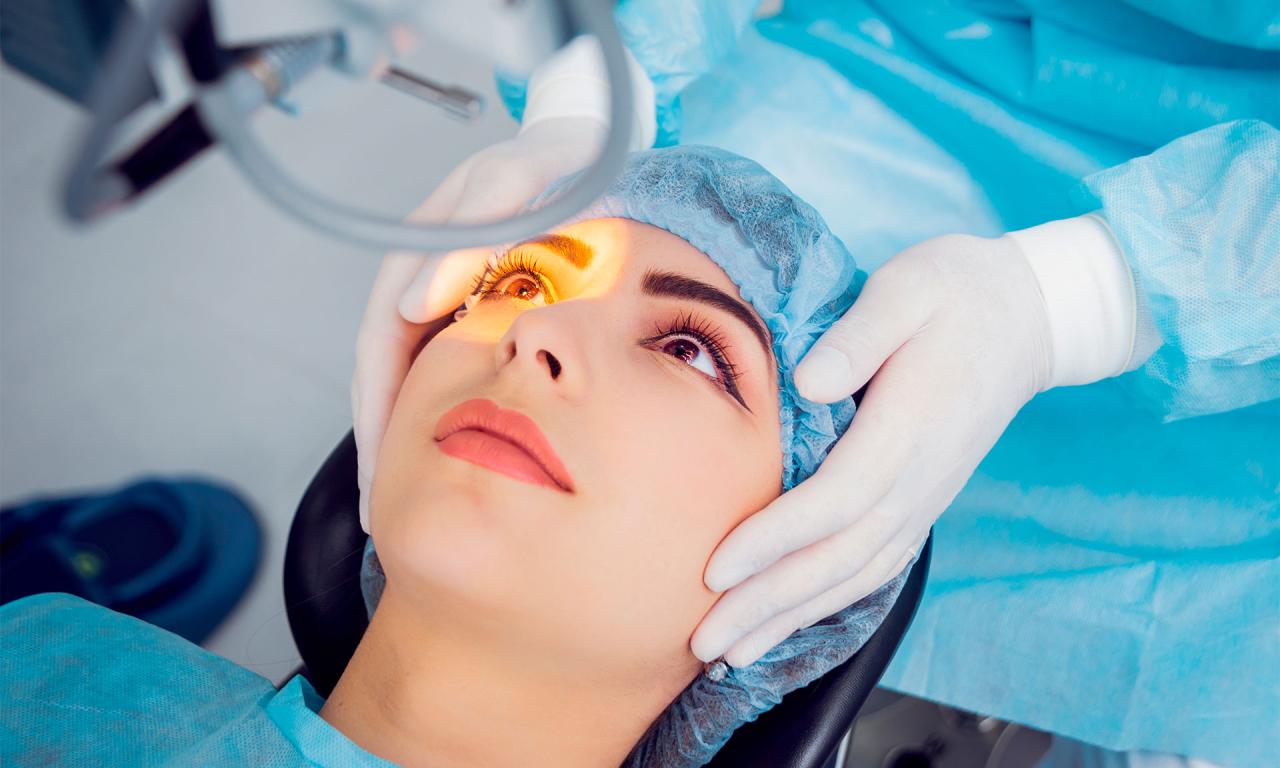
Refractive surgery involves reshaping the cornea, the transparent outer layer of the eye, to alter the way light enters the eye and focuses on the retina. This procedure is typically performed using a laser, but other methods also exist.
Laser-Assisted In Situ Keratomileusis (LASIK)
LASIK is a widely used refractive surgery technique. It involves creating a thin flap in the cornea, lifting it, and using an excimer laser to reshape the underlying corneal tissue. The flap is then repositioned, allowing the cornea to heal naturally. LASIK is effective for correcting myopia, hyperopia, and astigmatism.
Photorefractive Keratectomy (PRK)
PRK is another common refractive surgery technique that involves removing the outer layer of the cornea, the epithelium, before reshaping the underlying corneal tissue with an excimer laser. The epithelium regenerates over time, typically within a few weeks. PRK is an alternative for individuals who are not suitable for LASIK, such as those with thin corneas.
Intralase Femtosecond Laser
Intralase femtosecond laser is a technology used in LASIK and other refractive surgeries. It creates the corneal flap with precision and accuracy, minimizing the risk of complications. This technology allows for a more predictable and safer procedure.
Excimer Laser
Excimer lasers are used in refractive surgeries like LASIK and PRK to reshape the cornea. They emit ultraviolet light that removes corneal tissue in a precise and controlled manner. The laser is guided by a computer, ensuring accuracy and consistency.
Lens Implants
Lens implants are artificial lenses surgically placed within the eye to correct refractive errors. They are an alternative to refractive surgery, particularly for individuals with high refractive errors or who are not suitable for corneal surgery.
Intraocular Lenses (IOLs)
IOLs are small, artificial lenses implanted within the eye’s natural lens capsule. They are typically used to correct presbyopia, the age-related loss of near vision, and cataracts, clouding of the natural lens.
Multifocal IOLs
Multifocal IOLs have multiple focal points, allowing the eye to focus at different distances. This type of IOL can reduce or eliminate the need for reading glasses.
Toric IOLs
Toric IOLs are designed to correct astigmatism, a refractive error caused by an unevenly shaped cornea. They are typically used in cataract surgery to improve visual acuity.
Phacoemulsification
Phacoemulsification is a surgical procedure used to remove the natural lens during cataract surgery. It involves using an ultrasonic device to break up the lens into small pieces, which are then aspirated from the eye.
Contact Lenses
Contact lenses are thin, curved discs placed directly on the eye’s surface to correct refractive errors. They offer a non-surgical alternative to glasses, providing clear vision without the need for surgery.
Soft Contact Lenses
Soft contact lenses are made from flexible materials that conform to the shape of the eye. They are generally comfortable to wear and are available in various materials and designs.
Rigid Gas Permeable (RGP) Contact Lenses
RGP contact lenses are made from a rigid, gas-permeable material that provides sharper vision than soft lenses. They are typically used for individuals with astigmatism, irregular corneas, or other conditions that cannot be corrected with soft lenses.
Contact Lens Materials
Contact lenses are made from various materials, including silicone hydrogel, polyhema, and RGP materials. Each material has its own properties, affecting lens comfort, oxygen permeability, and durability.
Future Advancements in Vision Correction
The field of vision correction is constantly evolving, with ongoing research and development aimed at improving existing technologies and exploring new approaches.
Gene Therapy
Gene therapy holds promise for treating genetic eye disorders that affect vision. It involves delivering genes to the eye to correct defective genes or introduce new genes to restore vision.
Bioengineered Lenses
Researchers are exploring the development of bioengineered lenses that can be implanted in the eye to correct refractive errors. These lenses would be made from biocompatible materials and would integrate seamlessly with the eye’s natural tissues.
Artificial Corneas
Scientists are working on creating artificial corneas that can be transplanted to restore vision in individuals with corneal blindness. These artificial corneas would be biocompatible and would function like the natural cornea.
Teleophthalmology and Remote Monitoring
Teleophthalmology is a rapidly growing field that uses technology to deliver eye care services remotely. It plays a crucial role in expanding access to eye care, particularly in underserved areas and for patients who face challenges in accessing traditional healthcare facilities.
Technologies Used for Remote Patient Monitoring
Teleophthalmology leverages various technologies to facilitate remote patient monitoring. These technologies enable healthcare providers to assess patients’ conditions, diagnose eye diseases, and manage treatment plans remotely.
- Telemedicine Platforms: These platforms provide secure and HIPAA-compliant communication channels for virtual consultations, allowing patients to connect with ophthalmologists from anywhere with an internet connection. Video conferencing, messaging, and file sharing capabilities facilitate real-time interactions and data exchange.
- Wearable Devices: Smartwatches and other wearable devices equipped with sensors can collect valuable data on patients’ vital signs, activity levels, and sleep patterns. This information can be used to monitor patients’ overall health and identify potential eye-related issues early on.
- Remote Imaging: Teleophthalmology relies on remote imaging technologies, such as fundus cameras, OCT scanners, and tele-retinoscopes, to capture high-quality images of the eye. These images are transmitted securely to ophthalmologists for analysis and diagnosis.
Challenges and Opportunities Associated with Teleophthalmology
While teleophthalmology offers significant benefits, it also presents challenges that need to be addressed.
- Patient Privacy and Data Security: Teleophthalmology involves the transmission of sensitive patient information, which necessitates robust security measures to protect privacy and prevent data breaches. Secure data storage, encryption protocols, and access control mechanisms are crucial for maintaining patient confidentiality.
- Regulatory Frameworks: The adoption of teleophthalmology requires clear regulatory frameworks to ensure quality of care, patient safety, and compliance with ethical guidelines. Establishing standards for telemedicine practices, provider qualifications, and data privacy is essential for the responsible integration of teleophthalmology into healthcare systems.
- Accessibility and Equity: Teleophthalmology has the potential to bridge healthcare gaps and improve access to eye care for underserved populations. However, disparities in internet access, digital literacy, and affordability can limit the reach of teleophthalmology services. Addressing these challenges through initiatives that promote digital inclusion and affordable access to technology is crucial for ensuring equitable access to eye care.
Artificial Intelligence and Machine Learning in Ophthalmology

The integration of artificial intelligence (AI) and machine learning (ML) into ophthalmology has ushered in a new era of advancements, revolutionizing how eye diseases are diagnosed, treated, and managed. These technologies leverage the power of algorithms and data analysis to enhance diagnostic accuracy, personalize treatment plans, and ultimately improve patient outcomes.
Image Analysis and Disease Detection, Ophthalmology technology
AI and ML algorithms are adept at analyzing vast amounts of medical image data, including fundus photographs, optical coherence tomography (OCT) scans, and fluorescein angiography images. This capability allows for the automated detection of subtle patterns and anomalies that might escape the human eye, aiding in the early identification of various eye diseases.
- Diabetic Retinopathy: AI-powered algorithms can analyze fundus images to identify microaneurysms, hemorrhages, and other signs of diabetic retinopathy, enabling early detection and intervention, thus preventing vision loss.
- Glaucoma: ML models can analyze OCT scans to detect subtle changes in the optic nerve and retinal nerve fiber layer, which are early indicators of glaucoma, potentially leading to earlier diagnosis and treatment.
- Age-Related Macular Degeneration (AMD): AI algorithms can analyze fundus images to identify drusen, pigment changes, and other signs of AMD, facilitating early detection and monitoring of disease progression.
Treatment Planning and Surgical Assistance
AI and ML are also transforming treatment planning and surgical procedures in ophthalmology. These technologies can analyze patient data, including medical history, imaging results, and other relevant factors, to generate personalized treatment recommendations and predict potential complications.
- Cataract Surgery: AI-powered systems can assist surgeons in planning cataract surgery by predicting the optimal lens power and intraocular lens (IOL) type, leading to more accurate refractive outcomes.
- Glaucoma Surgery: ML algorithms can analyze patient data and OCT scans to predict the optimal surgical approach for glaucoma, minimizing complications and maximizing treatment efficacy.
- Retinal Detachment Surgery: AI-assisted surgical navigation systems can guide surgeons during retinal detachment surgery, enhancing precision and minimizing surgical errors.
Ethical Considerations and Challenges
While the potential benefits of AI in ophthalmology are immense, it is crucial to acknowledge the ethical considerations and challenges associated with its implementation.
- Data Privacy: Ensuring the privacy and security of patient data is paramount, as AI algorithms rely on vast amounts of sensitive medical information. Robust data protection measures are essential to safeguard patient confidentiality.
- Algorithmic Bias: AI algorithms are trained on data sets, and if these datasets contain biases, the algorithms may perpetuate those biases, potentially leading to unfair or inaccurate diagnoses and treatment recommendations.
- Human Oversight: While AI can assist in decision-making, it is essential to maintain human oversight and ensure that AI systems are used responsibly and ethically. Human expertise and judgment remain crucial in the interpretation of AI-generated results and the ultimate decision-making process.
Emerging Trends and Future Directions
The field of ophthalmology is constantly evolving, with new technologies emerging to revolutionize patient care, research, and our understanding of eye health. This section explores some of the most promising trends that are shaping the future of ophthalmology.
Bioprinting and Regenerative Medicine
Bioprinting, a revolutionary technology, holds immense promise for ophthalmology. It involves using 3D printing techniques to create biological structures, such as tissues and organs, from living cells. In ophthalmology, bioprinting is being explored for:
- Creating artificial corneas: Bioprinting could be used to create artificial corneas for patients with corneal diseases, such as keratoconus or Fuchs’ dystrophy. This could potentially eliminate the need for corneal transplants, which are currently limited by donor availability.
- Regenerating retinal cells: Bioprinting could be used to create new retinal cells for patients with age-related macular degeneration (AMD) or retinitis pigmentosa. This could potentially restore vision in patients who have lost sight due to these diseases.
- Developing personalized therapies: Bioprinting could be used to create personalized therapies for eye diseases. For example, a patient’s own cells could be used to create a customized patch for treating macular degeneration.
The potential impact of bioprinting on ophthalmology is vast. It could lead to new treatments for currently incurable diseases, reduce the need for invasive surgeries, and improve the quality of life for millions of people. However, there are challenges that need to be addressed, such as:
- Ensuring the biocompatibility of bioprinted tissues: Bioprinted tissues must be compatible with the patient’s body to avoid rejection.
- Developing efficient and scalable bioprinting techniques: Bioprinting techniques need to be efficient and scalable to produce enough bioprinted tissues to meet the needs of patients.
- Overcoming regulatory hurdles: Bioprinting is a relatively new technology, and regulatory approval for bioprinted tissues will be crucial for their widespread adoption.
Nanotechnology in Ophthalmology
Nanotechnology involves the manipulation of materials at the atomic and molecular level. This technology has the potential to revolutionize ophthalmology by providing new ways to diagnose, treat, and prevent eye diseases.
- Nanoparticles for drug delivery: Nanoparticles can be used to deliver drugs directly to the target tissues in the eye, improving treatment efficacy and reducing side effects. For example, nanoparticles can be used to deliver anti-VEGF drugs for the treatment of macular degeneration.
- Nanomaterials for diagnostic imaging: Nanomaterials can be used to enhance diagnostic imaging techniques, such as OCT and fluorescein angiography. This can help to improve the early detection and diagnosis of eye diseases.
- Nanobots for targeted therapy: Nanobots are tiny robots that can be programmed to perform specific tasks, such as delivering drugs or repairing damaged tissues. Nanobots are being explored for the treatment of glaucoma, diabetic retinopathy, and other eye diseases.
The potential impact of nanotechnology on ophthalmology is significant. It could lead to more effective and targeted treatments, improved diagnostic accuracy, and earlier intervention for eye diseases. However, there are challenges that need to be addressed, such as:
- Ensuring the safety of nanomaterials: Nanomaterials need to be carefully tested to ensure their safety for use in the eye.
- Developing efficient and targeted delivery methods: Nanomaterials need to be delivered to the target tissues in the eye in a safe and effective manner.
- Overcoming regulatory hurdles: Nanomaterials are a relatively new technology, and regulatory approval will be crucial for their widespread adoption.
Personalized Medicine in Ophthalmology
Personalized medicine involves tailoring medical treatment to the individual patient based on their genetic makeup, lifestyle, and other factors. This approach is becoming increasingly important in ophthalmology, as it can help to:
- Identify patients at risk for developing eye diseases: Genetic testing can be used to identify patients who are at an increased risk for developing certain eye diseases, such as AMD or glaucoma. This can allow for early intervention and potentially prevent vision loss.
- Predict treatment response: Genetic testing can be used to predict how patients will respond to different treatments. This can help to ensure that patients receive the most effective treatment for their individual needs.
- Develop personalized therapies: Personalized therapies can be developed based on the patient’s unique genetic profile. This can lead to more effective and targeted treatments with fewer side effects.
The potential impact of personalized medicine on ophthalmology is substantial. It could lead to more effective treatments, improved patient outcomes, and a better understanding of eye diseases. However, there are challenges that need to be addressed, such as:
- Ensuring the privacy and security of genetic data: Genetic data is sensitive and needs to be protected from unauthorized access.
- Developing cost-effective personalized therapies: Personalized therapies can be expensive to develop and implement.
- Overcoming ethical concerns: There are ethical concerns about the use of genetic information in healthcare, such as the potential for discrimination.
Artificial Intelligence and Machine Learning in Ophthalmology
Artificial intelligence (AI) and machine learning (ML) are rapidly transforming the field of ophthalmology. These technologies can analyze vast amounts of data, identify patterns, and make predictions.
- Automated image analysis: AI and ML can be used to automate the analysis of retinal images, such as OCT scans and fundus photographs. This can help to improve the accuracy and efficiency of diagnosis and disease monitoring.
- Predictive modeling: AI and ML can be used to develop predictive models that can identify patients at risk for developing eye diseases. This can allow for early intervention and potentially prevent vision loss.
- Robotic surgery: AI and ML are being used to develop robotic systems for ophthalmic surgery. This can improve the precision and accuracy of surgery, leading to better outcomes for patients.
The potential impact of AI and ML on ophthalmology is enormous. It could lead to more accurate diagnoses, improved treatment outcomes, and a better understanding of eye diseases. However, there are challenges that need to be addressed, such as:
- Ensuring the accuracy and reliability of AI and ML algorithms: AI and ML algorithms need to be rigorously tested to ensure their accuracy and reliability.
- Addressing ethical concerns: There are ethical concerns about the use of AI and ML in healthcare, such as the potential for bias and discrimination.
- Developing user-friendly AI and ML tools: AI and ML tools need to be user-friendly and accessible to ophthalmologists.
| Emerging Trend | Technology | Potential Impact | Challenges |
|---|---|---|---|
| Bioprinting | 3D printing of biological tissues | New treatments for incurable diseases, reduced need for invasive surgeries, improved quality of life | Biocompatibility, scalability, regulatory approval |
| Nanotechnology | Manipulation of materials at the atomic and molecular level | More effective and targeted treatments, improved diagnostic accuracy, earlier intervention for eye diseases | Safety of nanomaterials, efficient delivery methods, regulatory approval |
| Personalized Medicine | Tailoring medical treatment to the individual patient | Identify patients at risk, predict treatment response, develop personalized therapies | Privacy and security of genetic data, cost-effectiveness, ethical concerns |
| Artificial Intelligence and Machine Learning | Automated data analysis, predictive modeling, robotic surgery | More accurate diagnoses, improved treatment outcomes, better understanding of eye diseases | Accuracy and reliability of algorithms, ethical concerns, user-friendliness |
Conclusion
As ophthalmology technology continues to evolve, we can anticipate even more groundbreaking advancements in the future. These innovations hold the promise of restoring vision, preventing blindness, and improving the quality of life for countless individuals. The future of eye care is bright, thanks to the relentless pursuit of technological solutions.
Ophthalmology technology is rapidly advancing, with new tools and techniques constantly emerging to improve patient care. For those in need of reliable and efficient repair services for their ophthalmology equipment, the 12th man technology service center is a valuable resource.
They provide expert support for a wide range of ophthalmology equipment, ensuring that clinics and hospitals can continue to provide high-quality eye care.


